Arthroscopy is a common surgical procedure in which a joint(arthro-) is examined with a small camera (-scopy). Arthroscopy gives doctors a clear picture of the inside of the knee. This helps them diagnose and treat knee problems.
Technological advances have led to high-definition displays and high-resolution cameras. These and other improvements have made arthroscopy a very effective tool in the treatment of knee problems. According to the American Orthopedic Society for Sports Medicine, more than million knee arthroscopies are performed worldwide each year.
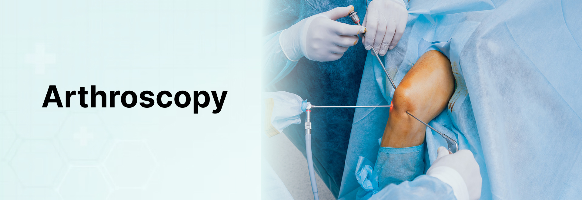
Arthroscopy is performed through small incisions. During the procedure, the orthopedist places an arthroscope (a small camera instrument, about the size of a pencil) into your knee joint. The arthroscope sends the image to the television. The surgeon can see the knee structures in great detail on the screen.
A surgeon may use arthroscopy to repair or remove damaged tissue. To do this, small surgical instruments are inserted through other incisions around the knee.
Shoulder Arthroscopy
Shoulder arthroscopy is surgery that uses a small camera called an arthroscope to examine or repair tissue in or around the shoulder joint. The arthroscope is inserted into the skin through a small incision.
Description
The rotator cuff is a group of muscles and tendons that cover your shoulder joint. These muscles and tendons hold your arm in your ball and socket shoulder joint, and they help you move your shoulder in different directions. The tendons in the rotator cuff can tear when they are overused or Injured.
Most people receive general anesthesia before this surgery. This means you will be unconscious and unable to feel pain. Or, you may have regional anesthesia. Your arm and shoulder area will be numbed so that you do not feel any pain in this area. If you receive regional anesthesia, you will also be given medicine to make you very sleepy during the operation
First, your surgeon will examine your shoulder with the arthroscope Your surgeon will
Insert the arthroscope into your shoulder through a small incision The arthroscope is connected to a video monitor in the operating room.
Inspect all the tissues of your shoulder joint and the area above the joint – the cartilage, bones, tendons, and ligaments
Repair any damaged tissues. To do this, your surgeon will make 1-3 more small incisions and insert other instruments through them. A tear in a muscle, tendon, or cartilage will be fixed. Damaged tissue may need to be removed.